Organoid-based Discovery platform for Innovative Screening, Evaluation and Identification
Organoid-based drug efficacy and
toxicity evaluation solution
ODISEI ONC
Oncology
ODISEI PET
Companion Animals
ODISEI VIR
Infectious Diseases
ODISEI NEP
Renal Diseases
ODISEIGUT
Nutraceuticals
ODISEI CNS
Brain Diseases
ODISEI SKIN
Cosmetics
ODISEI HEP
Liver Diseases
ODISEI Spatial
Spatial Biology
Overview
ODISEI Solution precisely predicts the efficacy of rapidly evolving new materials
Discrepancies between animal testing and human results
The rise of animal ethics and the spread of alternative methods to animal testing
Astronomical costs
involved
& Low success rates
Challenges in
evaluating new
modality drugs
Providing the optimal evaluation solution to accurately assess advanced pharmaceuticals and
new materials without animal testing, enhancing the efficiency of drug development
Advantages of Organoid Model
Experience more precise, easier, faster, and reliable drug efficacy evaluation ODISEI Solutions for your successful drug discovery.
TypeSimilarity with
human organsPhysiological
complexityTime for
EstablishmentSuccess
rateMass
productionEthical
issue
3D OrganoidHighRelatively
complexWeeksHighPossibleNo
2D Cells
(PDC)LowSimpleDaysHighPossibleNo
PDX Model (PDX)HighComplexMonthsLowImpossibleYes
Strategies: Basic Research
Connecting genotypes to specific phenotypes
Cancer initiation and progression
Clonal evolution
Cancer stem cell characterization
Mechanism of drug response and resistance
Utilization Strategy for Each Stage of New Drug Development
Target Section
1. New target excavation and verification
Lead Finding,
Prioritization Optimization
2. New drug screening 3. Drug efficacy Evaluate and optimize
Phase I
4. Clinical trial Prediction of success rate
Phase II
Phase III
5. Clinical trial protocol development
6. Treatment prediction marker development
Organoid Establishment and Banking
Over 20 diverse organoids, including TME (Tumor Microenvironment) , enabling cutting-edge trends like Neoantigen and Immunotherapy applications
Skin & Hair
Cerebral
Midbrain
Lacrimal Gland
Salivary Gland
Adenoid
Tonsil
Lung
Heart
Liver
Stomach
Kidney
Intestine
Prostate Cancer
Hepatocellular Carcinoma
Breast Cancer
Pancreatic Cancer
Renal Cell Carcinoma
Endometrial Cancer
Head & Neck Cancer
Non-Small Cell Lung Cancer
Cholangiocarcinoma
Gastric Cancer
Colorectal Cancer
Ovarian Cancer
Available pre-clinical research
Donor-matched immune microenvironment
Various patient-derived organoid lineups
High-resolution data
(HCS, FACS, NGS, scRNAseq, multiflex IHC)
Find out how we can be your partner in providing ethical choices and exceptional results
Client
Therapeutic Approaches
Antibodies
Anti-Cancer Drugs
Vaccines
Small Molecules
Peptides
Nucleosides
Microbiome
Virus
Cosmetic Compounds
Proinflammatory-Cytokines
Proinflammatory-Chemokines
Disease Modeling
Oncology Virus
Oncology Skin
Oncology Gut
Cytotoxic T Cells
Macrophages
DCs
Tregs
NKs
CAFs
Skin
Intestinal
Liver
Kidney
Cardiac
Lung
Stomach
Brain
Read-outs
Morphology
Cell viability
Cell type composition
Gene expression
Mutation profiling
Immune cell profiling
Pathway analysis
Cell to cell interaction
Metabolism
Analysis
High-content Screening
Cell Viability
Fluorescence-sorting
Fluorescence-activated Cell Sorting
Confocal Microscopy
Immunohistochemistry
Immunofluorescence
Phenocycler-fusion
(Multiplex-IHC)
-CODEX, OPAL
Bulk-RNAseq
Single Cell RNAseq
Whole Genome Sequencing
MS/MS
Report
ODISEI ONC
Precision oncology solution replicating patient tumors and immune microenvironments for anti-cancer and immunotherapy evaluation
High-Fidelity Preclinical Models
Oncology utilizes 3D patient-derived organoids (PDOs) cultured directly from tumor biopsies.
These models preserve the original tumor’s genetic and phenotypic characteristics, providing a more clinically relevant system than traditional 2D cell lines
or animal models.
Extensive Organoid Biobanking
A comprehensive biobank of PDOs, derived from a wide range of cancer types, forms an expansive repository that supports robust screening efforts.
This resource enables the identification of drug candidates across diverse tumor profiles and informs strategic decision-making throughout the R&D pipeline.
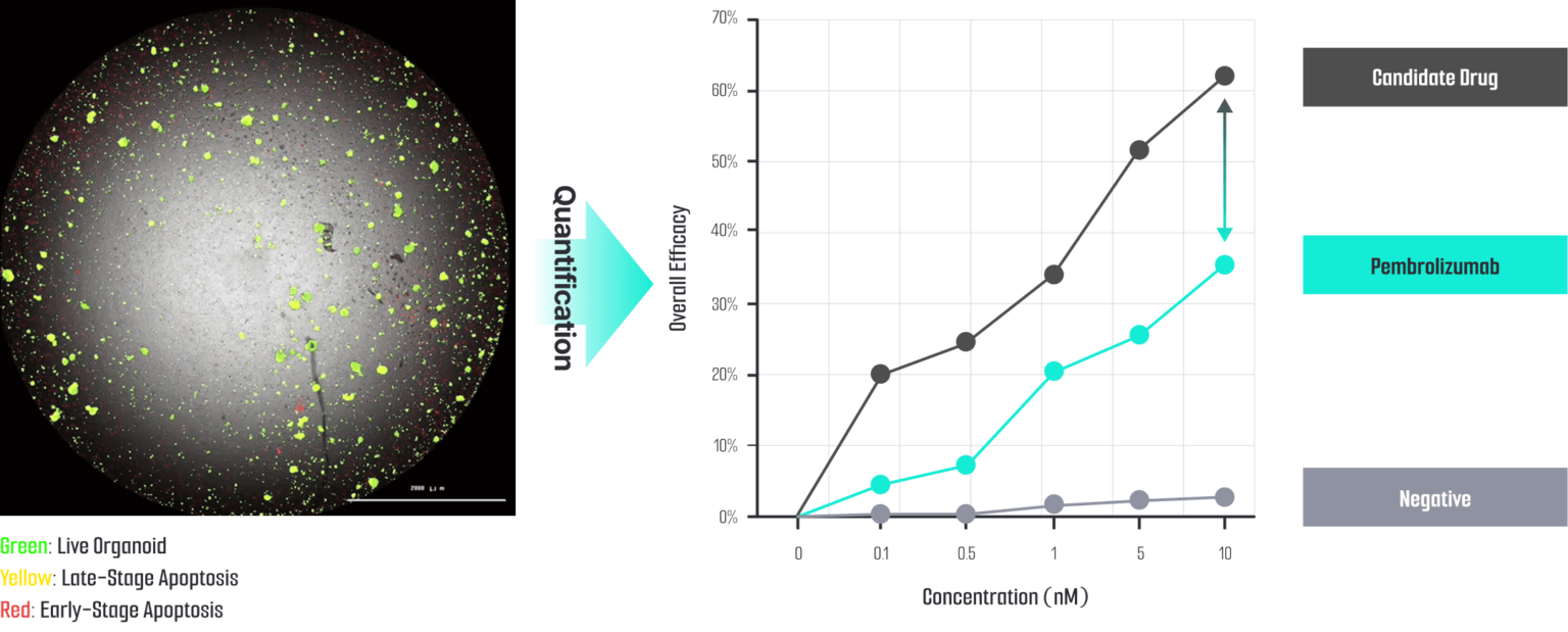
ODISEI-ONC : Immuno-Oncology Drug Evaluation Platform Using Tumor Organoids
Features
Enhance clinical predictive accuracy by developing advanced
models that more accurately recapitulate the Tumor Microenvironment (TME)
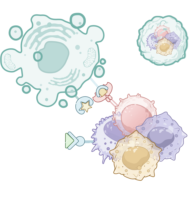
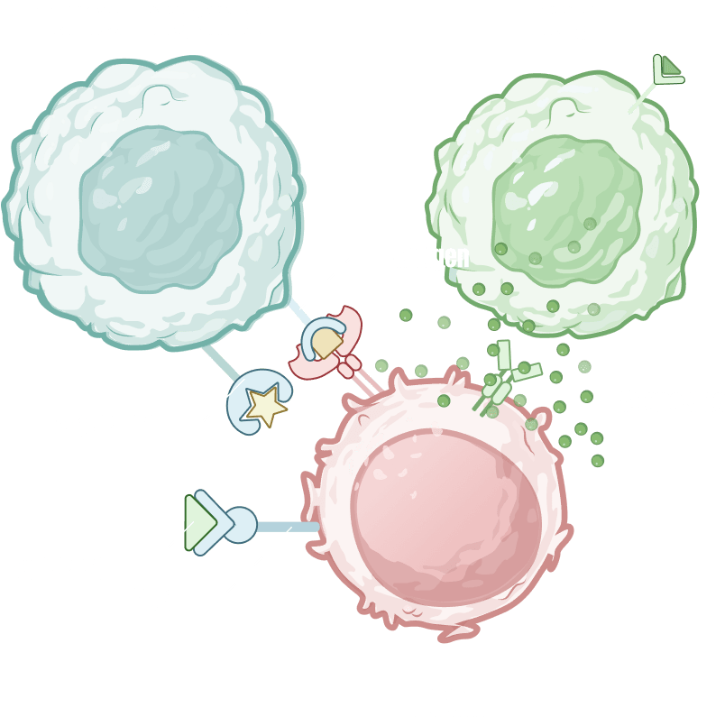
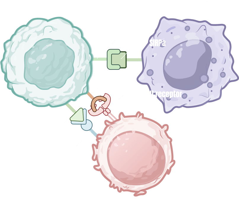
Immune-Microenvironment with Cytoxic T cells
Immune checkpoint determine whether cancer cells are killed after TCR-MHC binding
stablishment of an immune checkpoint blockade
evaluation platform utilizing cancer organoids and autologous T cells
Tumor-Infiltrating Lymphocyte (TIL)
Regulatory T cell (Treg)
Macrophage
Cancer Associate Fibroblast (CAF)
Validation Complete for caner organoid
Clinical information
Drug sensitivity test
Genetic mutation (WES analysis)
Specific cancer marker expression (Immunohistochemistry, Multiflex)
WES analysis in pancreatic cancer organoid
Drug sensitivity test in pancreatic cancer organoid
ODISEI GUT
Intestinal organoid-based solution enabling disease modeling and drug response prediction with organ-level functional fidelity
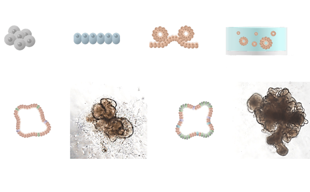
Generation of human intestinal organoid (hIO)
iPSC-derived intestinal organoids
iPSC-derived intestinal organoids are 3D models that replicate intestinal tissue using induced pluripotent stem cells (iPSCs), playing a key role in intestinal development and disease research. These organoids are generated by differentiating iPSCs into intestinal progenitor cells and cultivating them in a suitable extracellular matrix to form 3D structures. The organoids mature into structures with key intestinal layers and functions, which can be assessed through their absorption capacity, bacterial adhesion ability, and drug responses, mimicking real intestinal tissue.
Tissue-derived intestinal organoids
Tissue-derived intestinal organoids are 3D models created using intestinal cells directly isolated from tissue, providing a valuable tool for studying intestinal development, function, and diseases. These organoids are typically generated by culturing intestinal epithelial cells derived from intestinal tissue. Under specialized culture conditions, the cells form 3D structures that closely mimic the natural architecture and function of the intestine. By providing extracellular matrix (ECM) components and growth factors, the cells spontaneously organize into the key layers and morphology of the intestine, maintaining a microenvironment similar to that of the native tissue. These organoids are widely used for modeling intestinal diseases, drug testing, and studying both physiological and pathological conditions of the intestine.
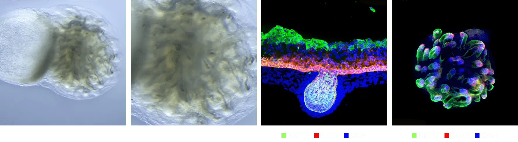
Cellular and structural similarity
The small intestine is a crucial organ responsible for nutrient absorption, consisting of four layers: the mucosa, submucosa, muscularis propria, and serosa. Inside the small intestine, villi maximize nutrient absorption, while crypts serve as regions where intestinal epithelial cells proliferate. Additionally, microvilli further expand the surface area for absorption.
The major cell types include enterocytes responsible for nutrient uptake, goblet cells that secrete mucus, Paneth cells that produce antimicrobial peptides, enteroendocrine cells that release hormones, and intestinal stem cells that regenerate the intestinal epithelium.
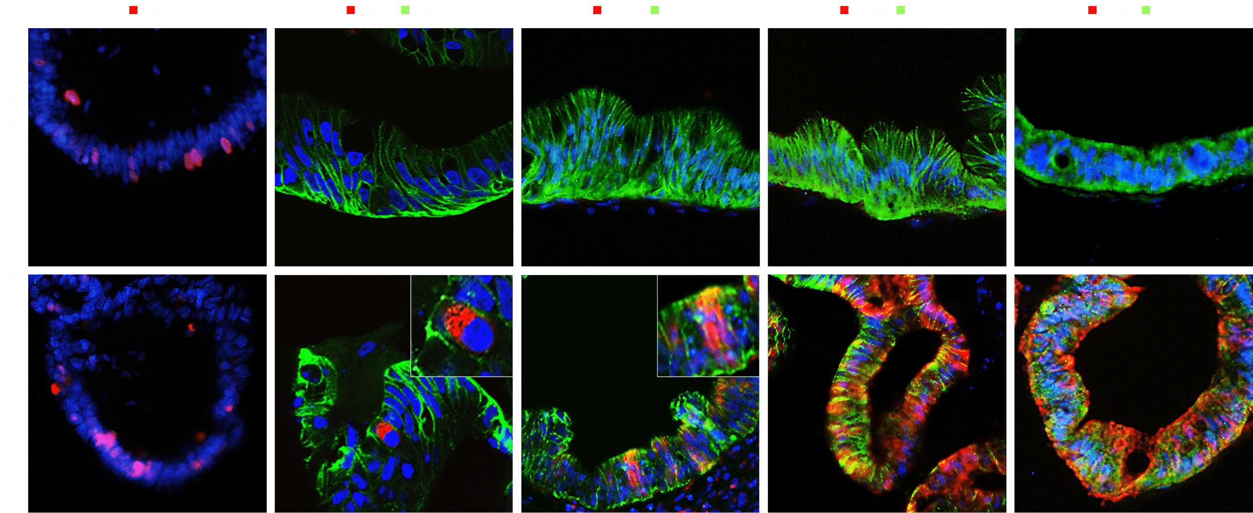
Intestinal organoids have a structure and cellular composition similar to human intestinal tissue and mimic the functions of the small intestine.
In our intestinal organoids, key markers for function and cell status have been identified, including Ki67, associated with cell proliferation; E-cadherin (Ecad), which maintains cell adhesion; OLFM4, a marker for intestinal stem cells; MUC13, involved in mucus secretion; and KRT20, indicative of mature intestinal epithelial cell differentiation.
Ki67 – Strongly expressed in proliferating cells within the crypts, indicating active cell proliferation
E-cadherin – Essential for maintaining adhesion between epithelial cells, ensuring structural integrity
OLFM4 – A key marker of intestinal stem cells, specifically expressed in the crypt region
MUC13 – Expressed in goblet cells and some enterocytes, contributing to the protection of the intestinal
KRT20 – Expressed in mature enterocytes, serving as an indicator of epithelial cell differentiation
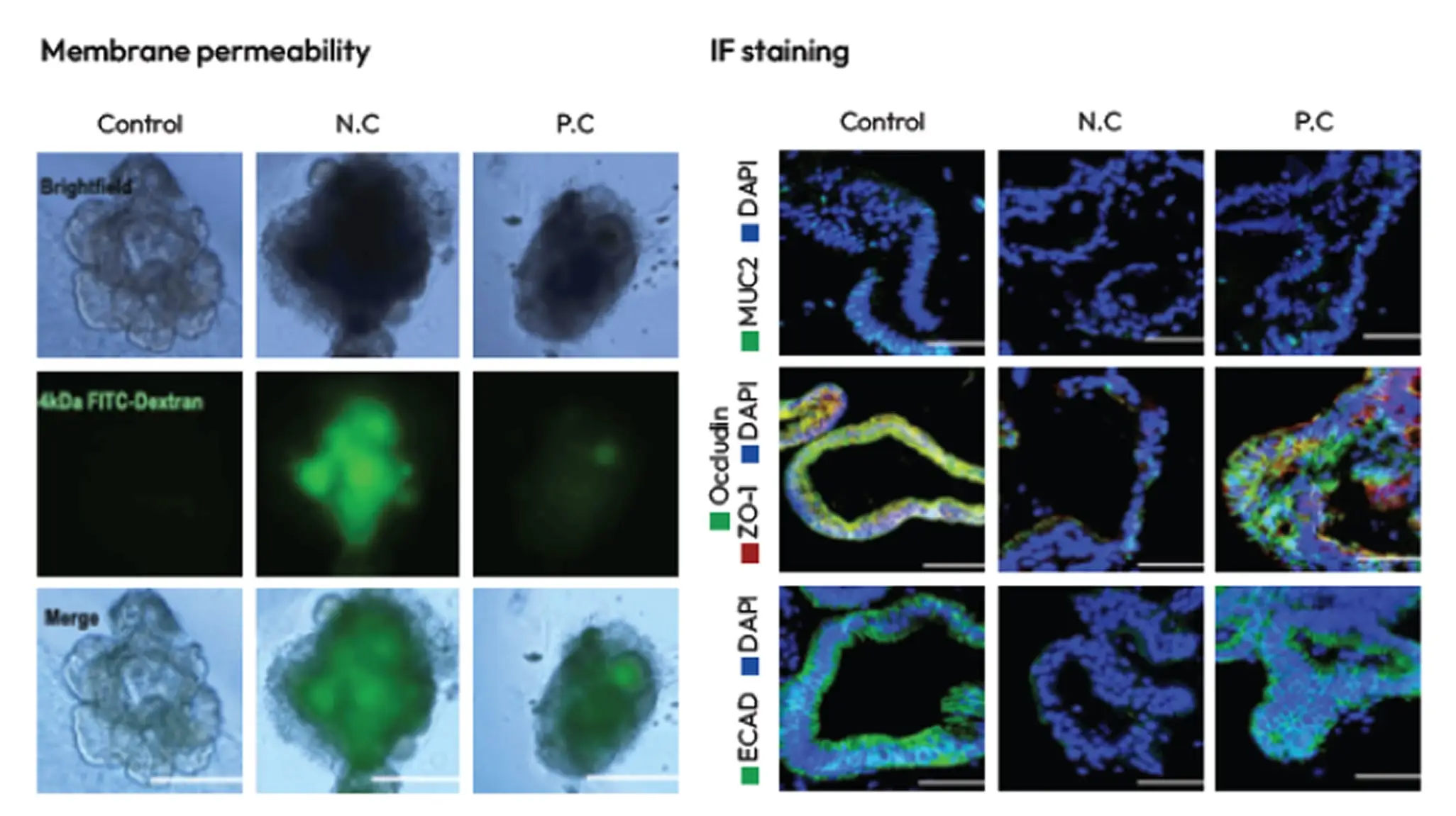
Intestinal Disease & Function Models
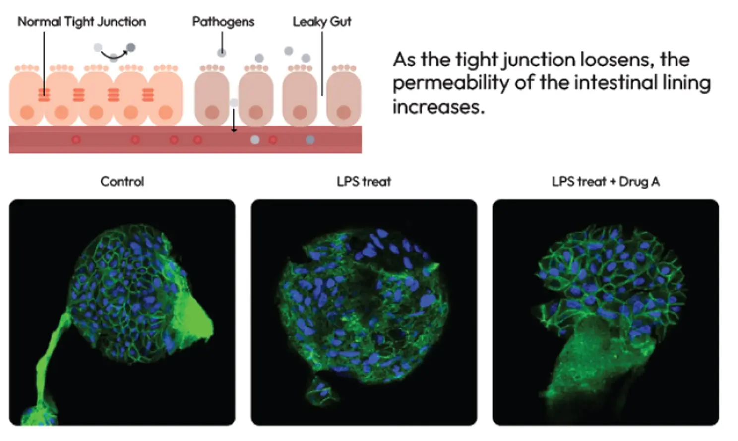
LGS (Leaky Gut Syndrome) Model
Development of leaky gut model causing diverse intestinal disease and treatment probiotics product to recovery.
As the tight junction loosens, the permeability of the intestinal lining increases
IBD (Inflammatory Bowel Disease) Model
Induction of Inflammatory Bowel Disease (IBD) model using Pro-Inflammatory cytokines and assessment of therapeutic efficacy.
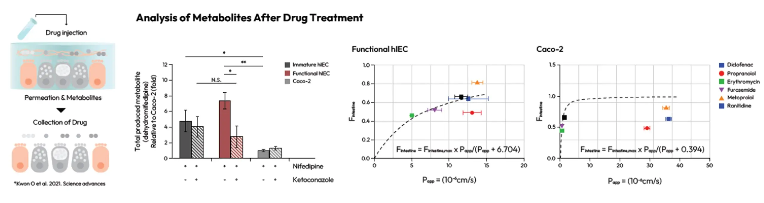
Drug Absorption and Permeability Evaluation Method
Results similar to human tests were obtained when evaluating drug metabolism and permeability in the intestinal epithelial model.
Analysis of Metabolites After Drug Treatment
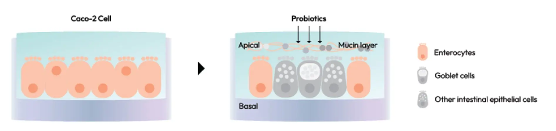
Microbiome & Probiotics
Discover Probiotic Potential with Our Gut-like hIESC Model Unlock the next generation of probiotic research.
Leverage our highly physiologically relevant hIESC-based intestinal model engineered to closely mimic
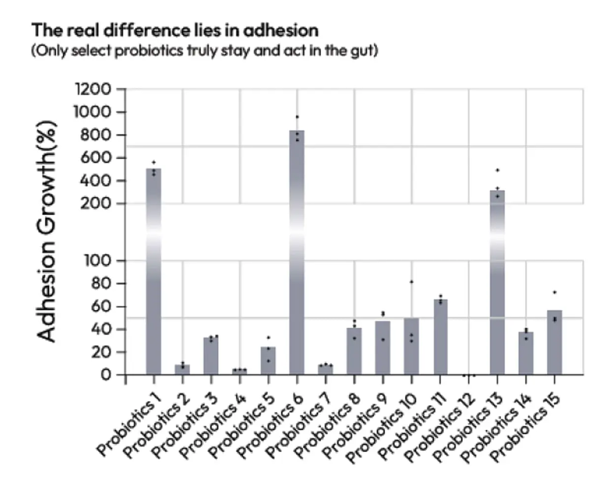
The real difference lies in adhesion
(Only select probiotics truly stay and act in the gut)
ODISEI SKIN
Human skin organoid solution recreating cellular structures and hair follicles for high-precision efficacy testing
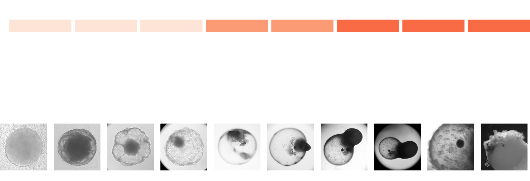
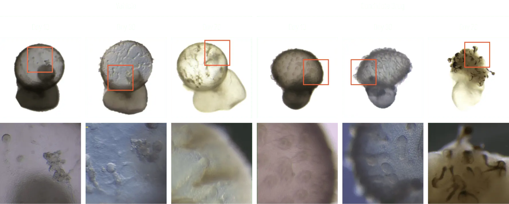
Generation of hair bearing skin organoid
We provide hair-bearing mature skin organoids developed through our differentiated technology based on hPSC-derived human skin organoids.
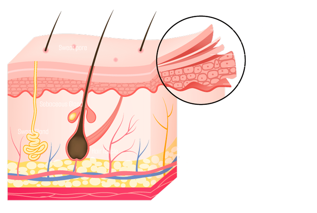
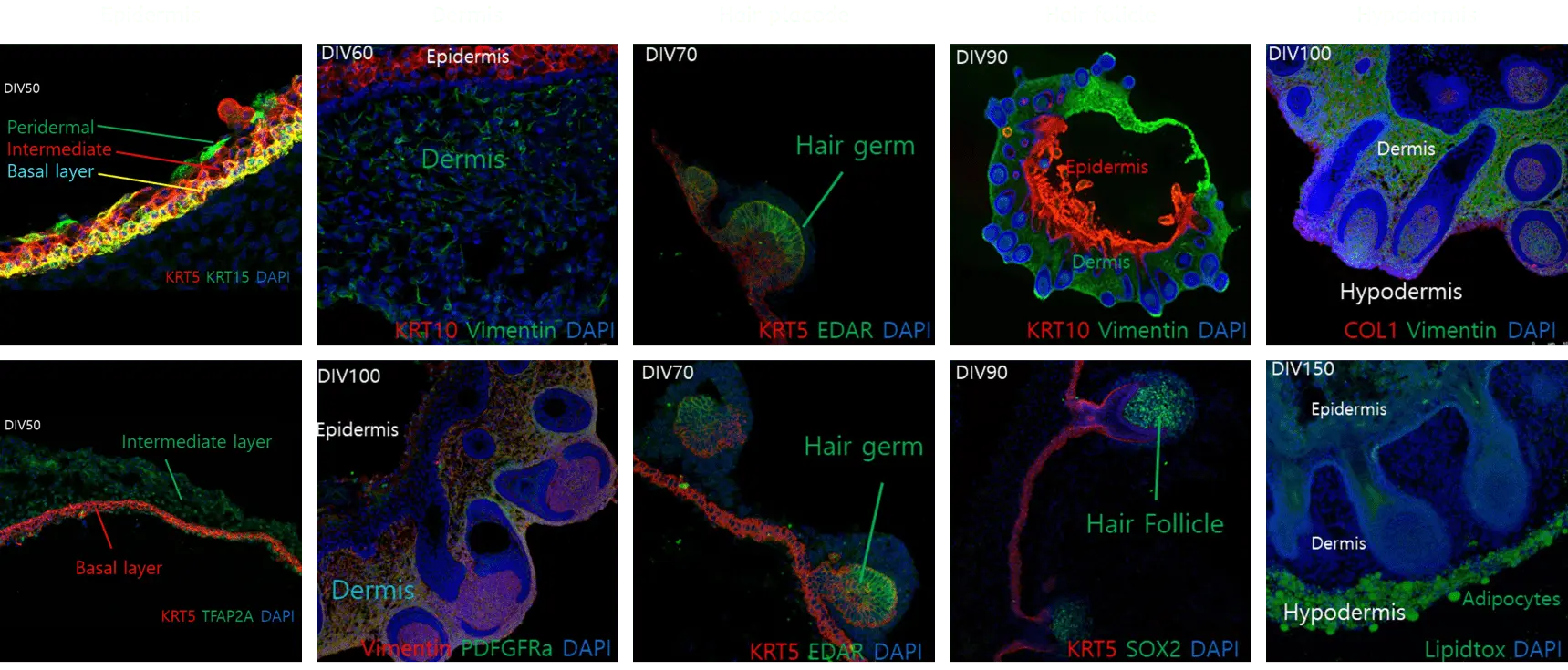
Cellular and structural similarity
Our skin organoids can be observed in a mature form with the epidermal layer, dermal layer, and subcutaneous tissue that appear in actual skin structure.
It is also composed of various cells such as fibroblasts, melanocytes, hair follicle-related cells, and adipocytes.
Structural characteristics of skin organoids
Skin Structure
Expression Markers
Epidermis
KRT5Basal Layer
KRT10Spinous Layer
KRT15Peridermal
TFAP2AIntermediate Layer
Dermis
VimentinFibroblast Intermediate Filament
PDFGFRαMesenchyme-
Associated Cell
COL1Collagen
EDARHair Germ
SOX2Hair Follicle
Hypodermis
LipidtoxAdipocytes
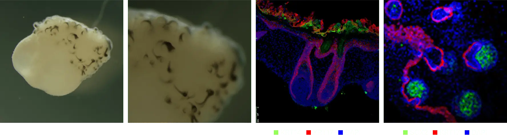
Advanced skin organoid manufacturing technology
Previous method
Our skin organoids can be observed in a structurally mature form and are capable of uniformly forming a higher number of hair follicles. Compared to traditional artificial skin models and 2D cell lines, our skin organoids exhibit superior performance in replicating the complexity and functionality of human skin.
Hypodermis
KRT5
KRT17
EDAR
SOX2
Skin structure
Basal Layer [Epidermis]
Cornified envelope [Epidermis]
Hair Gem [Dermis]
Dermal papilla cell [Dermis]
Advanced method
Evaluation of drugs for hair formation effectiveness
Structural characteristics of skin organoids
In order to discover drugs that help in the formation of hair, active substances were treated at the time when hair bulbs in skin organoids were formed, and drugs that increased hair follicle formation compared to the control group were selected.
When treated with an effective drug, an increase in the number of hair follicles was obtained, and additional research on mechanisms related to hair follicle formation was conducted.
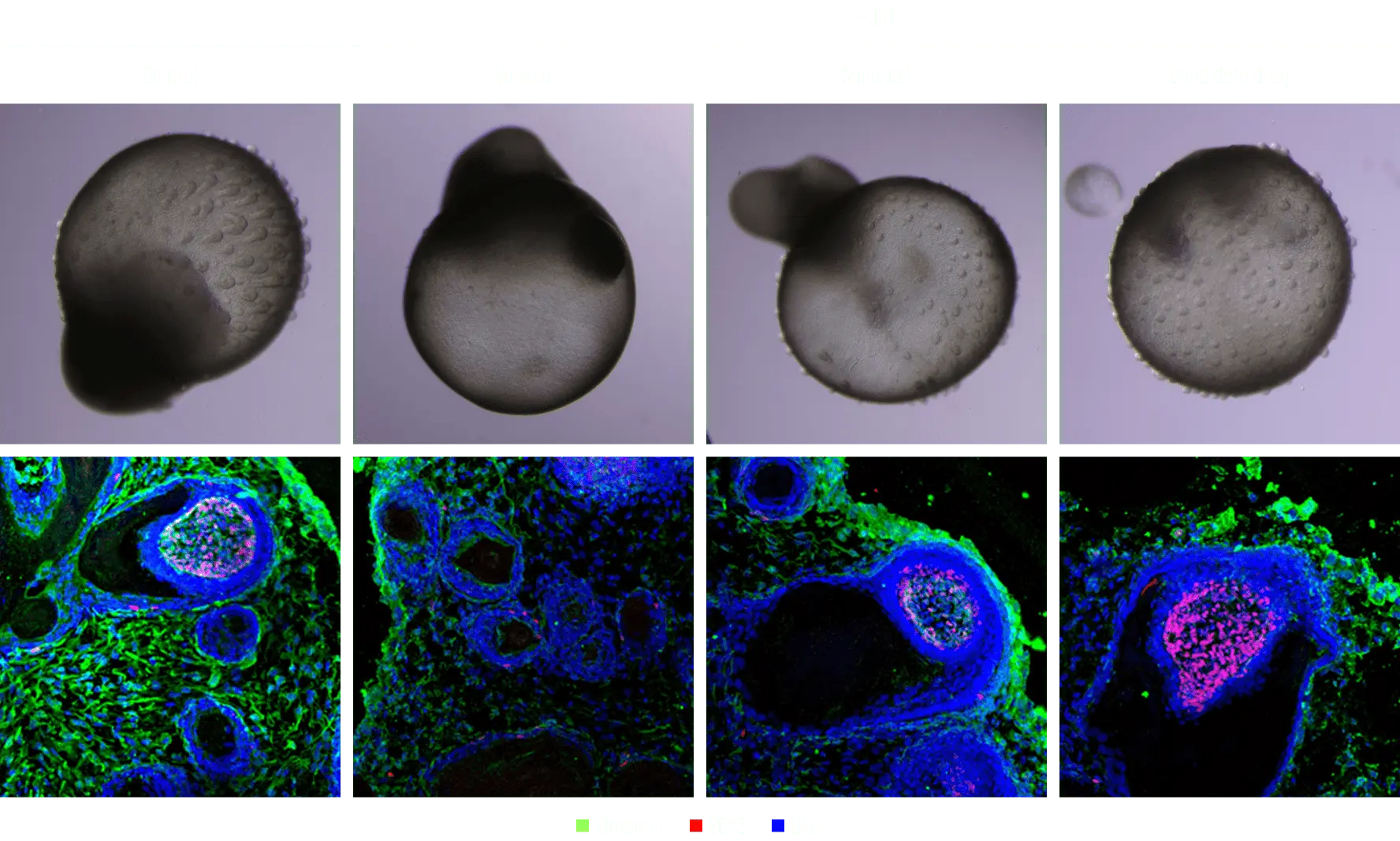
Hair formation efficacy test using skin organoids
Hair loss model production and effectiveness evaluation
To create a hair loss model, we treated skin organoids with hair with the male hormone DHT to create a hair loss model. It was confirmed that hair loss was suppressed by co-administration with Minoxidil, a known hair loss treatment, and was used to discover effective candidates.
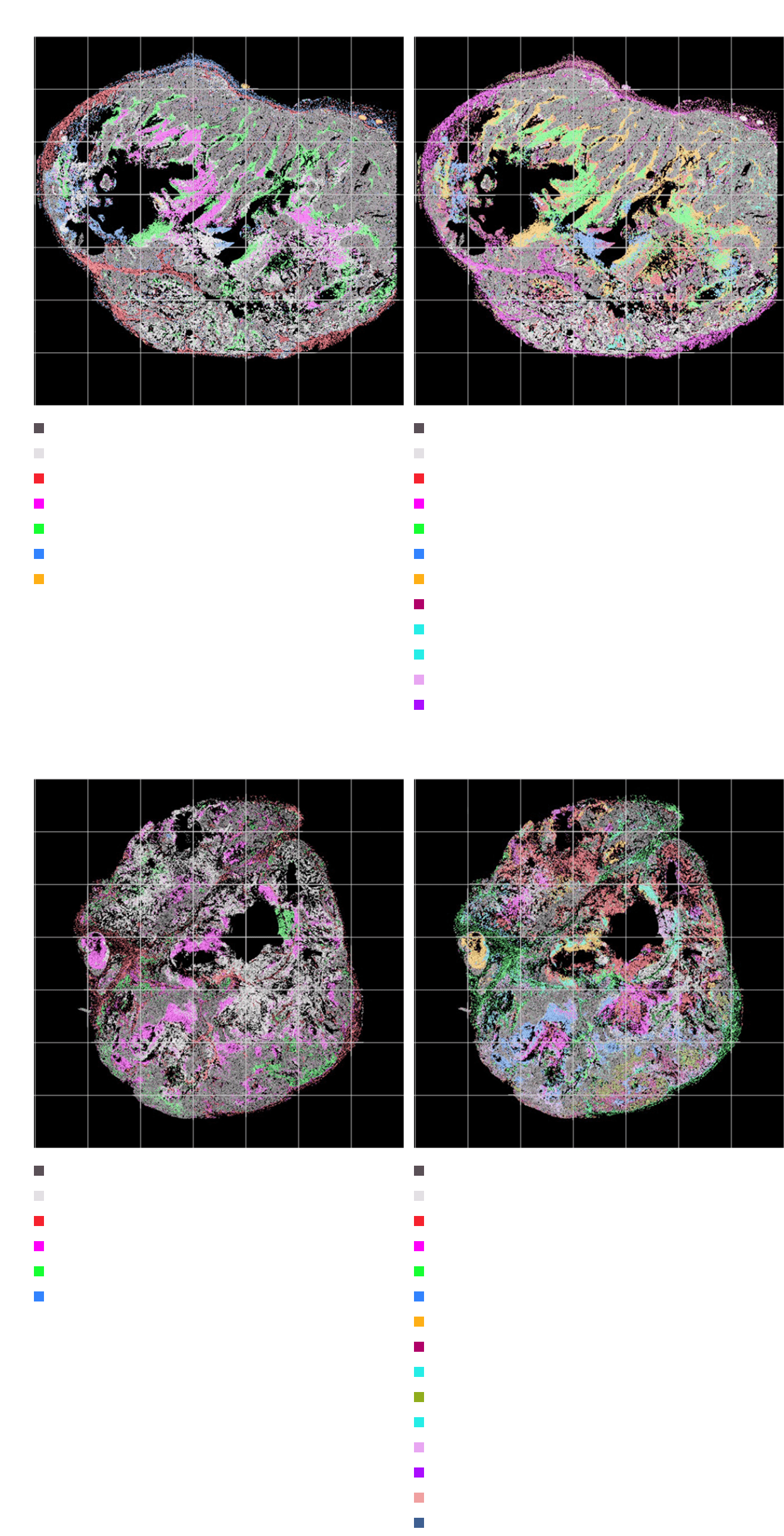
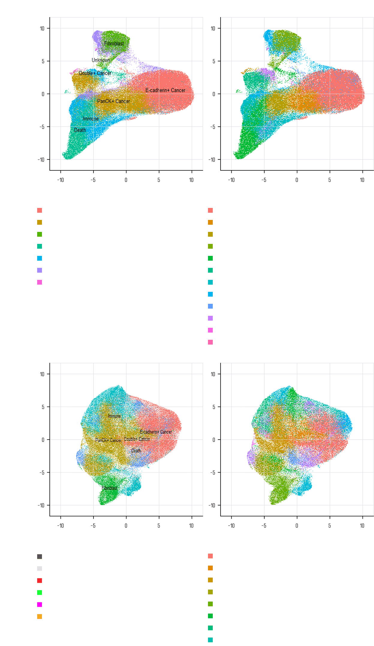
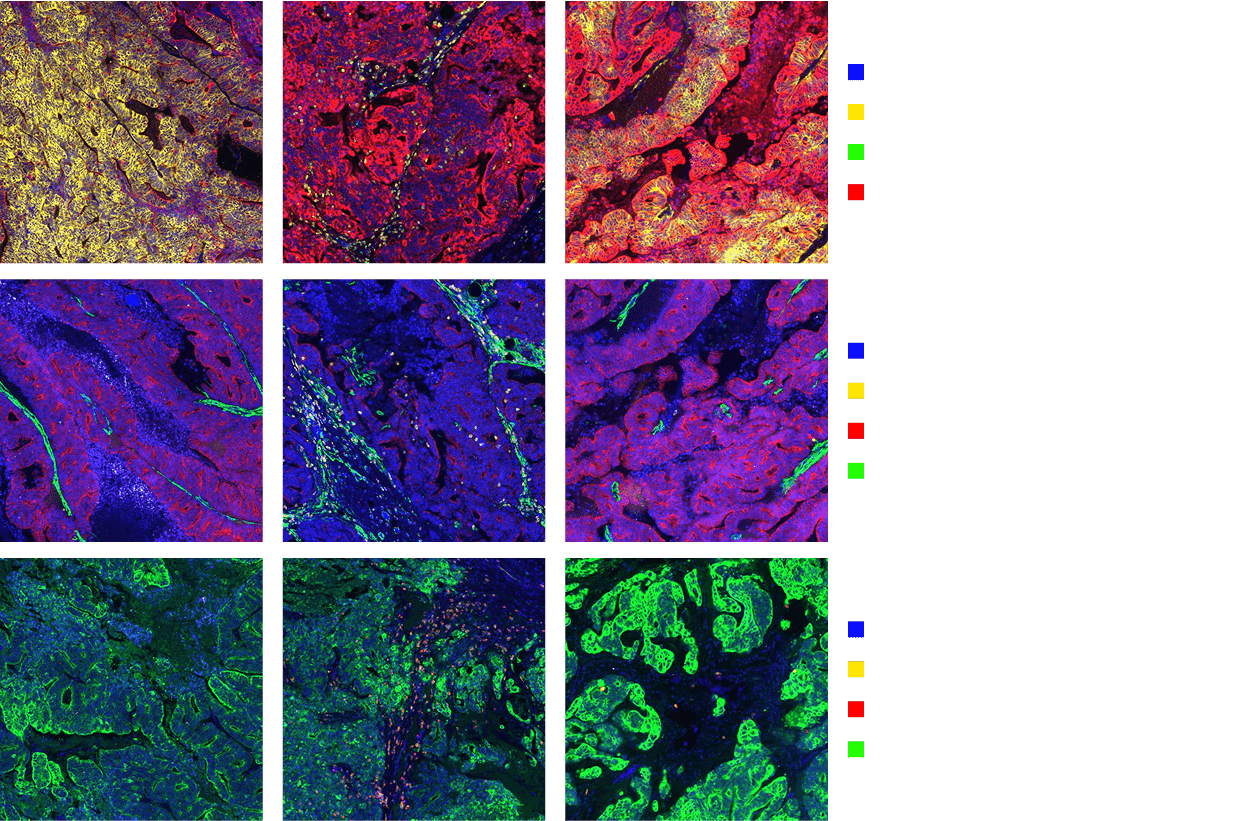
ODISEI Spatial
Spatial biology solution modeling tissue architecture and cell interactions for efficacy analysis and biomarker discovery
Phenocycler(CODEX)
CODEX was developed to overcome the limitations of existing IHC.
Multiplex-IHC is an experimental method that can confirm the expression of more than 100 biomarkers on one tissue slide.
It is possible to check the expression of various biomarkers on a single tissue slide and conduct comprehensive research on cell composition, cell function and state, and cell-cell interaction.
CODEX analysis is performed using DNA barcoding and uses antibodies conjugated to unique oligonucleotide sequences. By using a target specific barcode called dye-labelled reporter, it is possible to stain more than 100 biomarkers by
repeating the labeling cycle by dyeing three colors of fluorescence in one cycle.
Our Core Values
Staining more than 100 biomarkers on tissue slides 1 chapter.
Mapping of Millions of Cells Through Diverse Panel Combinations.
Single cell resolution analysis.
Cell phenotyping using marker staining combinations.
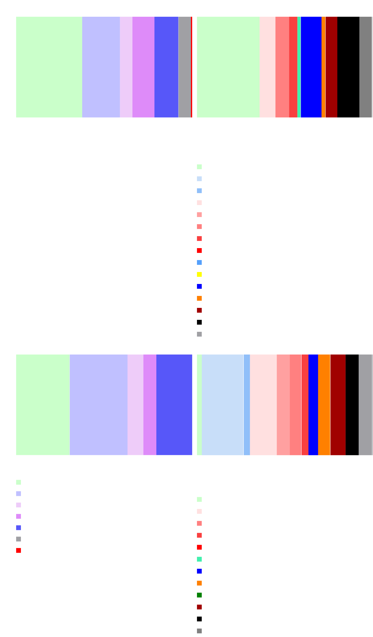
Cellular Neighborhood Analysis Utilizing Spatial Information of Cells.
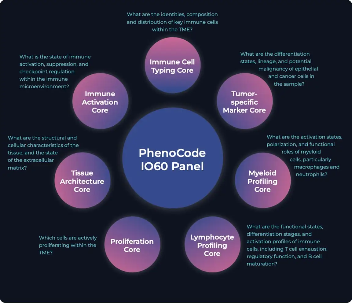
A new standard for Immuno-oncology research has arrived
Whether you’re looking to unlock the intricacies of the tumor microenvironment(TME), shed light on checkpoint therapy mechanisms, or drive breakthroughs in immunotherapy research, the PhenoCodeTM Discovery IO60 Human Protein Panel is a comprehensive, fast and scalable solution for ultrhigh-plex immune phenotyping-setting a new standard for immuno-oncology(IO) research.
New panel (IO60) Open
Advantage of IO60
Expanded panel enables deeper profiling of immune-tumor interactions.
Improved resolution in identifying rare immune subsets and tumor heterogeneity.
Enhanced capability for co-expression and spatial marker mapping.
More markers, more insight—accelerate biomarker discovery and therapeutic evaluation.
Immune cell type core
1. CD68
2. CD4
3. CD44
4. CD45RO
5. CD45
6. CD11c
7. CD8
8. HLA-A
9. HLA-DR
10. CD14
11. CD20
12. CD3e
13. CD56
Immune activation
1. IFNG
2. IDO1
3. PD-1
4. ICOS
5. PD-L1
6. LAG-3
7. VISTA
Tissue architecture
1. CD31
2. E-cadherin
3. SMA
4. Vimentin
5. CD34
6. Beta-actin
7. Podoplanin
8. Collagen IV
9. Caveolin
10. B-catenin1
Lymphocyte profiling
1. CD107
2. CD57
3. FOXP3
4. CD21
5. CD38
6. Granzyme B
7. TOX
8. TCF-1
9. CD79a
10. CD39
Myeloid profiling
1. iNOS
2. CD66
3. MPO
4. CD163
5. CD11b
6. CD206
7. CD209
Proliferation
1. Histone H3 Phospho(Ser28)
2. PCNA
3. Ki67
Tumor-specific marker
1. Keratin 14
2. SOX2
3. Pan-CK
4. Keratin 8/18
5. ER
6. Bcl-2
7. EpCAM
8. GP100
9. TP63
10. Keratin 5
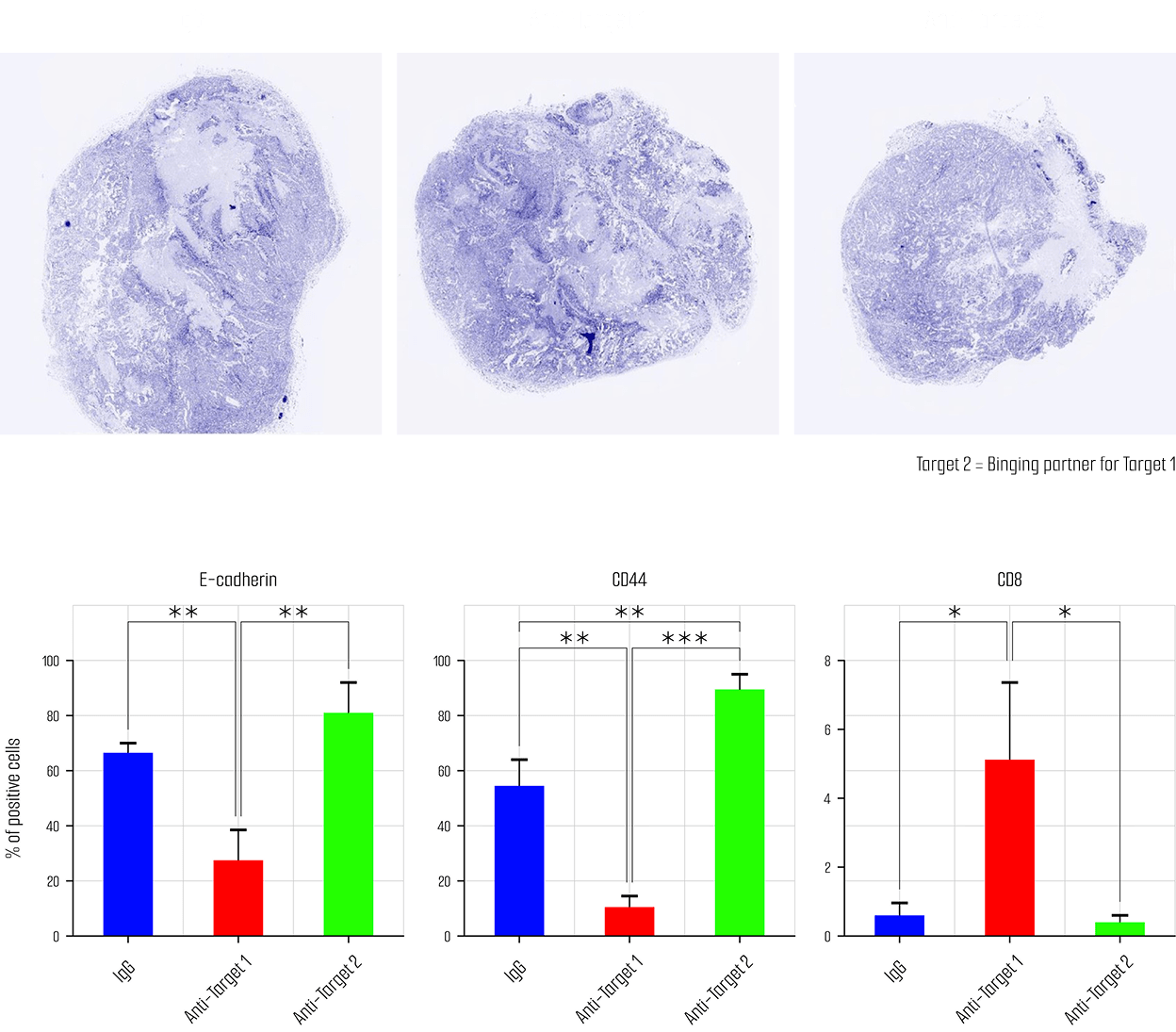
The published paper using our platform
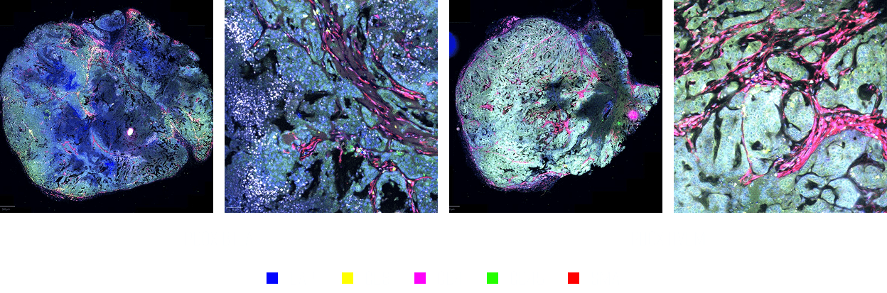
Read out
31markers (charged slide)
To identify the mechanism of a new target, acquire spatial information by simultaneously staining 31 marker in PDOX tissue.
Teratoma formation analysis
Simultaneous staining of 31 markers in tumor tissue
Identification of treatmentmechanism through spatial analysis of cell types
Identifying immunological mechanisms underlying the efficacy of anticancer immunotherapy and discovering biomarker candidates through theapplication of spatial biology
Toward a Sustainable Future with ORGANOIDSCIENCES
Join ORGANOIDSCIENCES in leading groundbreaking organoid research and the advancement of next-generation cell therapies.
Be part of the journey of biomedical innovation that is shaping the future of healthcare.