High-Plex Spatial Biology Analysis Services
Not just what cells express, but where they are located, how they interact, and why they respond differently to treatment.
ORGANOIDSCIENCES’ Spatial Biology Services provide high-plex single-cell spatial analysis to comprehensively characterize disease mechanisms, the tumor microenvironment (TME), and therapeutic responses.
Trusted Worldwide
Only
10
Qualified Providers Worldwide
Only
2
Providers in Asia
Only
1
Qualified Provider in Korea
Officially recognized by Akoya as a Qualified CRO Provider for high-plex spatial biology services.
- Official Akoya Qualification
- PhenoCycler™ Fusion Expertise
- Publication-Ready Spatial Analysis
- End-to-End Standardized Workflow
- 100+ Protein Multiplex imaging
Why Choose PhenoCycler™ Fusion for Spatial Biology?
Choose the Right Spatial Biology Technology for Your Research
-
FeaturePhenoCycler™ FusionMultiplex IFSpatial Transcriptomics
-
Primary TargetProteinProteinRNA
-
Spatial Context✓✓✓
-
Single-cell Resolution✓PartialNear Single-cell*
-
Multiplex Capability100+ proteins~8 proteinsThousands of genes
-
Best ApplicationCell Phenotyping Tumor Microenvironment (TME) Biomarker Discovery Drug Response AnalysisTargeted Protein ImagingGene Expression Profiling
How PhenoCycler™ Fusion Works
Discover More Than Expression
Go beyond protein expression to understand cellular location, phenotype, and cell-to-cell interactions through automated high-plex spatial protein imaging.
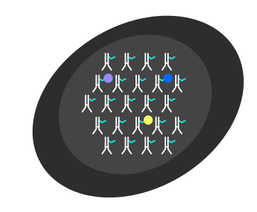
1DNA-Barcoded Antibody Technology
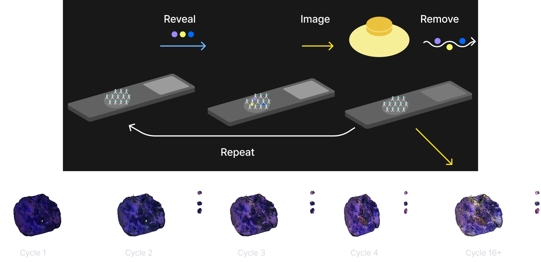
2Automated Reveal–Image–Remove Cycles
Automated reveal, image, and remove cycles continuously capture every biomarker with high reproducibility and minimal manual intervention
3High-Plex Spatial Mapping
End-to-end Workflow
Sample Submission
Staining
Imaging
Bioinformatics Analysis
Report Delivery
How Spatial Biology Accelerates Your Research
Transform Spatial Data into Biological Insight
Tumor Microenvironment
- Immune Cell Distribution
- Immune Hot / Cold Tumor
- Cell-to-Cell Interaction
- Tumor Heterogeneity
Target Validation
- Target Expression
- Tumor Specificity
- Spatial Localization
- Off-target Assessment
Drug Response
- Drug Response Prediction
- Mechanism of Action
- Resistance Analysis
- Treatment Response Evaluation
Biomarker Discovery
- Cell Phenotyping
- Co-expression Analysis
- Spatial Signature Discovery
- Novel Biomarker Identification

Explore Our Research Cases
Download real-world case studies and sample reports.
Explore publication-ready reports and analysis results from IO60™, Neuro42™, and Spatial Biology research projects.
Multiplex Protein Panels
60-marker human IO antibody panel
Immune cell type core
- CD68HLA-A
- CD4HLA-DR
- CD44CD14
- CD45ROCD20
- CD45CD3e
- CD11cCD56
- CD8
Lymphocyte Profiling
- CD107aGranzyme B
- CD57TOX
- FOXP3TCF-1
- CD21CD79a
- CD38CD39
Immune Activation
- IFNGPD-L1
- IDO1VISTA
- PD-1LAG3
- ICOS
Tissue Architecture
- CD31Beta-actin
- E-CadherinPodoplanin
- SMACollagen IV
- VimentinCaveolin
- CD34β-Catenin1
Proliferation
- Histone H3 Phospho (Ser28)
- PCNA
- Ki67
Tumor-Specific Markers
- Keratin 14Bcl-2
- SOX2EpCAM
- PanCKGP100
- Keratin 8/18TP63
- ERKeratin 5
Myeloid Profiling
- iNOSCD11b
- CD66CD206
- MPOCD209
- CD163
24-marker mouse FFPE IO antibody panel
T Cell Markers
- CD3eT cell
- CD4Helper T cell
- CD8Cytotoxic T cell
- FOXP3Regulatory T cell
- CD44Activated T cell
- CD45R/B220Naïve T cell
B Cell Markers
- CD20B cell
Structural, Proliferation and Tumor Markers
- Vimentin Mesenchymal cells, epithelial-mesenchymal transition (EMT)
- Col1A1Extracellular matrix
- CaveolinIntegral membrane protein
- PanCKEpithelial cells, tumor marker
- Ki67Proliferation marker
Other Cell Type Markers
- CD31Endothelial cells
- CD36Adipocyte progenitors
- Ter119Erythroid cells
Tumor-Specific Markers
- CD68 Macrophage
- CD206M2 macrophage
- F4/80Macrophage
Immune Cell Markers
- CD11cPanleukocyte
- Ly6gPeripheral neutrophils, monocytes and granulocytes
- FcRγMultiple immune cell types
- CD45Nucleated hematopoietic cells
- Iba1Microglia
- S100A9Myeloid differentiation
9-marker human metabolism antibody panel
Nutrient Uptake
- ASCT2GLUT1
PPP / Redox Balance
- G6PD
Glycolysis / Lactate Metabolism
- HK1LDHA
Mitochondrial Metabolism / OXPHOS
- CSIDH2
- SDHAATP5A
42-marker human neurobiology antibody panel
Neuronal Cell Type Markers
-
Mature NeuronsNeuN
MAP2
Neurofilament
Synaptophysin - GABAergic NeuronsGABA
- Cholinergic NeuronsCHAT
- Serotonergic NeuronsSerotonin
Neurodegenerative Disease Markers
-
Alzheimer's DiseaseAmyloid-beta
APOE
Tau (phospho T181)
Tau (phospho T231)
Tau (phospho S396)
Tau (phospho T205) - Parkinson's Disease & SynucleinopathiesAlpha-synuclein
- General NeurodegenerationH2A.X
Neuroplasticity & Cellular Remodeling
-
Proliferation & NeurogenesisPCNA
Ki67 -
Astrocyte & Structural PlasiticityS100B
Vimentin -
OligodendrocytesOlig-2
MBP
Blood-Brain Barrier Integrity & Vascular Components
-
Endothelial Cells & Vascular StructureClaudin-5
CD31
CD34
Collagen IV - Pericytes & Smooth Muscle CellsSMA
- Astrocyte-Endothelial InteractionsAQP4
Neuroinflammation & Immune Response
- MicrogliaTMEM119
-
Activated Microglia & MacrophagesIba1
CD68
Mac-2/Galectin-3
CD11b
F4/80
CD206 -
T cell
InfiltrationCD3e
CD4
CD8 -
General
Immune
ActivationCD45
CD44
CD20
MHC II
Ter119 -
Astrocyte- Associated
InflammationGFAP
42-marker mouse neurobiology antibody panel
Neuronal Cell Type Markers
-
Mature NeuronsNeuN
MAP2
Neurofilament
Synaptophysin - GABAergic NeuronsGABA
- Cholinergic NeuronsCHAT
- Serotonergic NeuronsSerotonin
Neurodegenerative Disease Markers
-
Alzheimer's DiseaseAmyloid-beta
APOE
Tau (phospho T181)
Tau (phospho T231)
Tau (phospho S396)
Tau (phospho T205) - Parkinson's Disease & SynucleinopathiesAlpha-synuclein
- General NeurodegenerationH2A.X
Neuroplasticity & Cellular Remodeling
-
Proliferation & NeurogenesisPCNA
Ki67 -
Astrocyte & Structural PlasiticityS100B
Vimentin -
OligodendrocytesOlig-2
MBP
Blood-Brain Barrier Integrity & Vascular Components
-
Endothelial Cells & Vascular StructureCD31
Claudin-5
Col1A1 - Pericytes & Smooth Muscle CellsSMA
- Astrocyte-Endothelial InteractionsAQP4
Neuroinflammation & Immune Response
- MicrogliaTMEM119
-
Activated Microglia & MacrophagesIba1
CD68
Mac-2/Galectin-3
CD11b
F4/80
CD206 -
T cell
InfiltrationCD3e
CD4
CD8 -
General
Immune
ActivationCD45
CD44
CD20
MHC II
Ter119 -
Astrocyte- Associated
InflammationGFAP
Elevate the Standard of Your Research
1. Unique Advantage for ADC Research
PhenoCycler-powered spatial profiling enables the simultaneous analysis of drug distribution and target expression within tissue.
In a single experiment, researchers can evaluate target expression, drug distribution, tumor heterogeneity, and spatial biomarker mapping—key factors for advancing ADC development.
Applications
✓ ADC Development
✓ Target Validation
✓ Companion Diagnostics
✓ Translational Research
2. Advanced Sample Processing
Key Benefits
✓ Organoid & 3D Tissue Expertise
✓ Reduced Experimental Failure Risk
✓ High-Quality Multiplex Imaging
3. End-to-End Spatial Analysis
Beyond delivering raw image data, our bioinformatics experts transform complex spatial datasets into publication-ready biological insights.
We provide comprehensive analytical outputs that enable researchers to interpret results and accelerate scientific discovery.
Deliverables
✓ Cell Phenotyping
✓ Cell-to-Cell Interaction Analysis
✓ Neighborhood Analysis
✓ Clustering & Spatial Mapping
✓ Publication-Ready Figures
✓ Comprehensive Analysis Report
4. Ready-to-Use Validated Panels
To reduce the time and cost required for panel development and validation, we provide pre-validated, research-ready multiplex panels.
Researchers can begin projects immediately with optimized panels designed for reproducible and reliable spatial profiling.
Available Panels
✓ IO60™ Immune-Oncology Panel
✓ Neuro42™ Neurobiology Panel
✓ Custom Panel Design Support
Key Benefits
✓ Faster Turnaround Time (TAT)
✓ Reduced Validation Cost
✓ Immediate Project Initiation
✓ Reproducible Results
5. Integrated Analysis Services Connected to the ODISEI™ Platform
Spatial Biology analysis can be integrated with the ODISEI™ Drug Evaluation Platform, allowing a single research project to be expanded into a comprehensive drug evaluation workflow.
Deliverables
✓ Drug efficacy assessment
✓ ADC evaluation
✓ Toxicity assessment
✓ Disease modeling
✓ Multi-omics analysis
6. Trusted Analysis Services for Researchers Worldwide
ORGANOIDSCIENCES supports the entire research process, from experimental execution and data analysis to the delivery of publication-quality results.
Deliverables
✓ High-quality data
✓ Publication-ready reports
✓ Expert bioinformatics analysis
✓ Dedicated scientific support
High-Plex Spatial Biology CRO Services
Toward a Sustainable Future with ORGANOIDSCIENCES
Be part of the journey of biomedical innovation that is shaping the future of healthcare.
Technical Inquiry
bd@organoidrx.com
PR/Marketing inquiry
pr@organoidrx.com
Recruitment inquiry
hr.admin@organoidrx.com
General Inquiry
info@organoidrx.com